

by Troy Vander Molen, CEO and doctor of physical therapy
 As a physical therapist with 20 years of clinical experience, I have seen a LOT of patients with lower back pain. In fact, it’s the most common condition for which my clients – and those of my teammates at Kinetic Edge – seek treatment on a daily basis.
Of the plethora of clients with which I’ve consulted, I wish I had a dime for every time I’ve heard:
- I don’t know why I hurt. I haven’t done anything different than normal recently.
- Don’t I need an MRI? How else will you know what to do?
My response to the first statement is nearly always the same. Though your lower back pain may come on suddenly, it is almost always a cumulative process that has been developing under the surface for years that eventually led to the pain you are experiencing now. The problem is that our movement dysfunctions – the limitations and compensations we make as a result of them – have occurred so gradually that they feel normal. We no longer know what it feels like to move ideally and efficiently.
Don’t get me wrong. I’m glad that the body can make compensations or we would experience pain far more often than we do now. However, the fact that we make compensations means that other areas often suffer as a result of that adjustment. Therefore, the real solution to your lower back problem is to find and fix the movement issue so that the area in the lower back that has experienced the physical stress no longer gets punished.
My response to needing an MRI is a bit more direct. No. In almost every case, you do not need an MRI. You probably don’t even need an X-ray. In fact, these internal pictures often provide you unnecessary information that is detrimental to your healing process and your pocketbook. Studies show that on average outcomes are worse and more costly for those who have these diagnostic tests early in the process.
Let’s take a quick look at the statistics and research behind MRIs specifically for disc herniation. Studies show that 60% of people that are given an MRI for any reason will have a disc bulge or disc herniation. The odds are slightly lower for younger people and slightly higher for older people. However, the presence of a disc bulge is hardly a rare occurrence regardless of your age. Despite its prevalence, of the 60% who have a diagnosed disc bulge, only 10% have pain!
Basically, what that means is that the MRI often times can actually confuse the provider and make someone think that they have a disc herniation or a disc bulge – which is completely accurate and objectively true by the way – without understanding and fixing what caused it and without even being assured that the pain is coming from that particular finding.
We also know that there is a big cost associated with MRIs – over $2,000 on average – and they are only valid for about a year. They just don’t help as much as most think they will. In fact, unless you are experiencing foot drop or loss of bowel/bladder continence, the results are unlikely to impact your treatment plan. Most physicians will still recommend conservative physical therapy care first.
There are health providers such as Physical Therapists, Orthopedic Surgeons, and Physiatrists that use simple and inexpensive special tests, which are movement or positional tests that will easily tell you the condition you’re suffering from. Research has shown that these simple tests have a high level of specificity and sensitivity and therefore can identify the source of lower back pain.
For instance, I can quickly determine if someone has stenosis in the lower back by giving them a verbal questionnaire which has been proven to be 97% accurate. The questionnaire includes three simple questions:
- Are you over the age of 55?
- Do you have lower back pain with standing and walking?
- Does your lower back pain go away with sitting?
If you answered “yes” to all 3 of these questions, then there is a 97% chance that you have lumbar stenosis. That has been proven by research, and it is more accurate than an MRI or an X-ray.

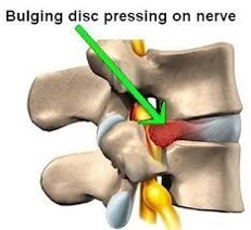
Lower back pain treatment experts also use special tests for disc herniations and disc bulges like a Straight Leg Raise or a Dural Stretch Test (which stretches the sciatic nerve). If those tests are positive, there is a 90% chance that the person has a disc herniation or a disc bulge.
We also determine that pretty quickly by simply asking the patient, do you have increased pain down your leg with coughing, laughing, or sneezing? If you do, that’s a very strong indicator that you have a disc herniation or disc bulge. This test is much more accurate – and much less expensive – than an MRI.
So, as you can see, the problem with these diagnostic tests is that, though they might tell you that you have stenosis, a disc bulge, or sacroiliac (SI) pain (which typically doesn’t show up on an MRI or an X-ray), the idea that an MRI or an X-ray is going to help the provider fix your lower back pain is usually false. There’s a quicker, simpler, and more cost-effective way to get the valuable information necessary to make you move better, feel better, and function better
The reason that I say this is because all three of those lower back conditions – stenosis, disc bulge, and SI pain – are a result of a different problem, something that we refer to as the root cause. The root cause is what is actually causing damage to the lower back.
 I like to use the analogy of an iceberg. What you see above the water is only 10% of the ice. Below the water is the other 90%. To understand lower back pain, you must realize that it is not the lower back’s fault that it has a disc herniation or SI inflammation or stenosis. There’s a bigger, deeper issue. It is the areas around the lower back that ultimately aren’t moving properly that shift the stress to another area, and the lower back is often the victim.
In fact, all of the conditions of lower back pain are ultimately a stress response. Yes, the lower back does have some damaged tissue. And yes, this damage is caused by physical stress. So yes, an MRI or X-ray helps identify which type of lower back pain you might have, but it doesn’t help you find the treatment or the solution for your lower back pain.
In my opinion, in most cases an X-ray or an MRI should be used as a pre-surgical tool. If all else fails and a surgeon is going to open you up, he needs to have an MRI or X-ray to determine what part of the spine he is going to surgically alter. You may have noticed that I didn’t use the word “fix” because the surgeon is almost always dealing with a secondary issue, not the root cause. Any and all lower back surgeries don’t fix the root cause. The surgery just alters the resulting problem that is caused by the root issue. The root issue is what caused the compensation and the cumulative stress that resulted in tissue damage at the other painful area.
I recently spoke with a physical therapy friend that practices in the beautiful northern Michigan town of Traverse City. Andrew Gorecki is a phenomenal physical therapist and a lower back expert. He had the opportunity to work with a local physician who had developed severe sciatica and lower back pain on the right side of his body. He was hardly able to walk. He happened to see Superior Physical Therapy’s advertisement in the local paper for a lower back pain and sciatica workshop, and he showed up the night of the event.
After the workshop, the physician came up to Andrew to tell him his personal lower back pain story. The physician had recently gone to his primary care physician who offered him pain medications for his symptoms, but he was struggling because he wasn’t legally allowed to take pain medications during his work hours. So, he had recently gotten an MRI which showed he had a disc bulge on the left side of his spine, and he immediately consulted with a neurosurgeon who recommend he have surgery immediately. In fact, at the time of the workshop, he was scheduled to have a surgery the following week.
His question to Andrew was this: “How can a disc bulge on the left side of my spine cause pain down my right leg and in my right lower back?” The answer Andrew gave him was simple: It can’t.
The physician was actually relieved with that answer. Since he was a physician, he was slightly embarrassed that he didn’t know the answer or believe the neurosurgeon expert. But deep down inside he knew that it was not plausible, and something didn’t feel right about surgery.
So, after speaking with Andrew, the physician chose to postpone his surgery and committed to 15 physical therapy visits. Every visit he was able to stand a little straighter and walk with less pain. Eventually he was pain free, moving well, and functioning normally again without the need for pain medications or surgery.
The reason the physician was able to find success was because Andrew’s physical therapy team found and treated his root problem. The client’s real issue was a loss of hip mobility – a pretty common finding, by the way – which was causing increased stress in his lower back. His disc bulge was only a secondary development that wouldn’t have gotten better with surgery unless he found a way to solve the real, primary problem.
I often have patients who come in with their imaging results. Sometimes they tell me that they aren’t sure if their symptoms fit with the image they have in their hand. If you are one of these people, don’t worry about it. You’re not alone. Move forward and seek to find the solution to your problem.
If you are one of those people who think you need an MRI or X-ray, please reconsider. Unless you have one of a very few major findings, it is an unnecessary expense. You can be confident in avoiding the diagnostic testing because the research shows that what the image shows you may be very misleading. Trust me, I have seen this happen hundreds and hundreds of time in the past. I’m here to tell you that the MRI and the X-ray often lie to you. These tests are not the most accurate way to identify lower back pain, and they are very unlikely to help you find your solution.
An MRI or X-ray does not tell anyone what to do about it, what the cause is, or what the treatment should be. That image is simply another tool, one that should be used only when somebody is about to have surgery so that the surgeon knows the exact level to go to when he is going to alter the structure of the lower back.
